
Under a microscope, a drop of blood turns out to be a crowd of cells. On a stained slide, you’ll see thousands of red blood cells as small pink discs with pale centers and no nucleus. Scattered among them are a few much larger white blood cells with dark purple nuclei, and tiny purple specks called platelets. The liquid part, plasma, doesn’t show up as anything at all; it’s the background the cells float in.
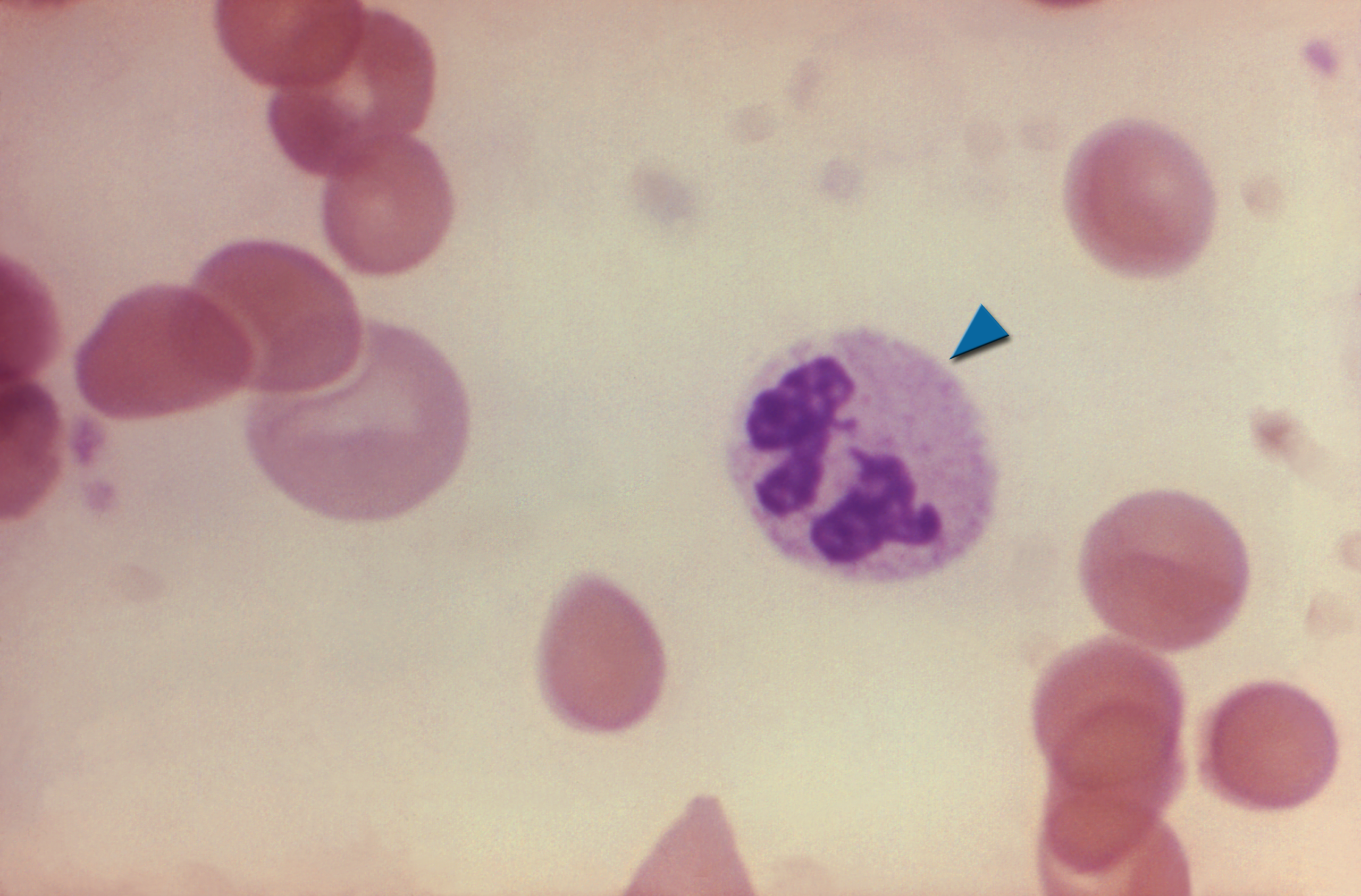
This guide shows what each part of blood looks like, what you’ll see at each magnification, and how to look at blood yourself safely.
Blood components at a glance
| Component | What it looks like on a stained slide | Size | How common | Main job |
|---|---|---|---|---|
| Red blood cells (erythrocytes) | Pink discs with a pale center, no nucleus | 6–8 µm | By far the most numerous cells: about 40–45% of blood volume | Carry oxygen and carbon dioxide |
| White blood cells (leukocytes) | Larger cells with a purple-stained nucleus | 7–20 µm depending on type | Roughly 1 for every 600–700 red cells | Fight infection |
| Platelets (thrombocytes) | Tiny purple fragments between the red cells | 2–3 µm | About 1 for every 10–20 red cells | Help blood clot |
| Plasma | Not visible on a slide; pale yellow liquid in a tube | n/a | About 55% of blood volume | Carries water, proteins, nutrients and waste |
To see blood this way you need a compound microscope with magnification up to 400x or, ideally, 1,000x. A stereo microscope or a pocket microscope can’t resolve individual blood cells. New to the controls? See our labeled guide to the parts of a microscope.
What blood looks like at different magnifications
| Total magnification | What you’ll see |
|---|---|
| 40x (4x objective) | A thin pink film; individual cells are too small to make out. Use this to find a well-spread area of the smear. |
| 100x (10x objective) | A carpet of tiny pink dots (red cells), with the occasional purple speck (a white cell’s nucleus). |
| 400x (40x objective) | Red cells are clearly round discs with pale centers. White cells stand out, and you can see the shape of their nuclei. |
| 1,000x (100x oil immersion) | The full detail: lobes of the white cell nuclei, granules in their cytoplasm, and individual platelets. |
Red blood cells (erythrocytes)
Red blood cells are the most numerous cells in blood. Under the microscope, they look like small round discs, pink on a stained slide, with a paler center. That pale center, called central pallor, appears because each cell is a biconcave disc, thinner in the middle than at the edges, like a doughnut without the hole. Each one is about 6–8 µm across.
Mature human red blood cells have no nucleus and no mitochondria. Losing the nucleus leaves more room for hemoglobin, the iron-rich protein that binds oxygen and gives blood its red color. Red cells carry oxygen from the lungs to the rest of the body and bring carbon dioxide back to the lungs to be breathed out.
In a well-made smear, red cells sit side by side without overlapping. In a thick area of the slide, they pile up and can stack like coins, which makes them harder to see individually.
Can you see blood type under a microscope?
No. Blood types depend on antigens, molecules on the surface of red blood cells, which are far too small to see with a light microscope. Type A blood carries A antigens, type B carries B antigens, type AB carries both, and type O carries neither. The Rh factor (positive or negative) is another surface antigen. Blood type is found with a lab test that mixes blood with antibodies and checks whether the cells clump together.
White blood cells (leukocytes)
White blood cells are part of the immune system. They’re larger than red blood cells but far fewer, roughly one for every 600–700 red cells. Unlike red cells, they have a nucleus, and that nucleus is what makes them easy to spot on a stained slide: it stains dark purple.
On their own, white cells are almost colorless, so blood is stained before viewing. Common stains like Wright’s or Giemsa color the nuclei purple and the granules in different cells pink, orange or dark blue. That’s how the five types are told apart.
The five types fall into two groups. Granulocytes have visible granules in their cytoplasm, and agranulocytes have clear cytoplasm.
| Type | Group | Share of white cells | Size | How to recognize it |
|---|---|---|---|---|
| Neutrophil | Granulocyte | 40–70% (most common) | 10–12 µm | Nucleus with 2–5 lobes joined by thin strands; fine, pale lilac granules |
| Lymphocyte | Agranulocyte | 20–40% | 7–15 µm | Round, dark nucleus that fills most of the cell; thin rim of blue cytoplasm |
| Monocyte | Agranulocyte | 2–8% | 12–20 µm (largest) | Kidney- or horseshoe-shaped nucleus; gray-blue cytoplasm, sometimes with small holes (vacuoles) |
| Eosinophil | Granulocyte | 1–4% | 12–15 µm | Two-lobed nucleus; large bright orange-red granules |
| Basophil | Granulocyte | Under 1% (rarest) | 10–14 µm | Dark blue-purple granules that often hide the nucleus |
Neutrophils
Neutrophils are the most common white blood cells and the body’s first responders to bacterial infection. They engulf and digest bacteria. They’re easy to recognize by their segmented nucleus, usually 2–5 lobes linked by thin strands.
Lymphocytes
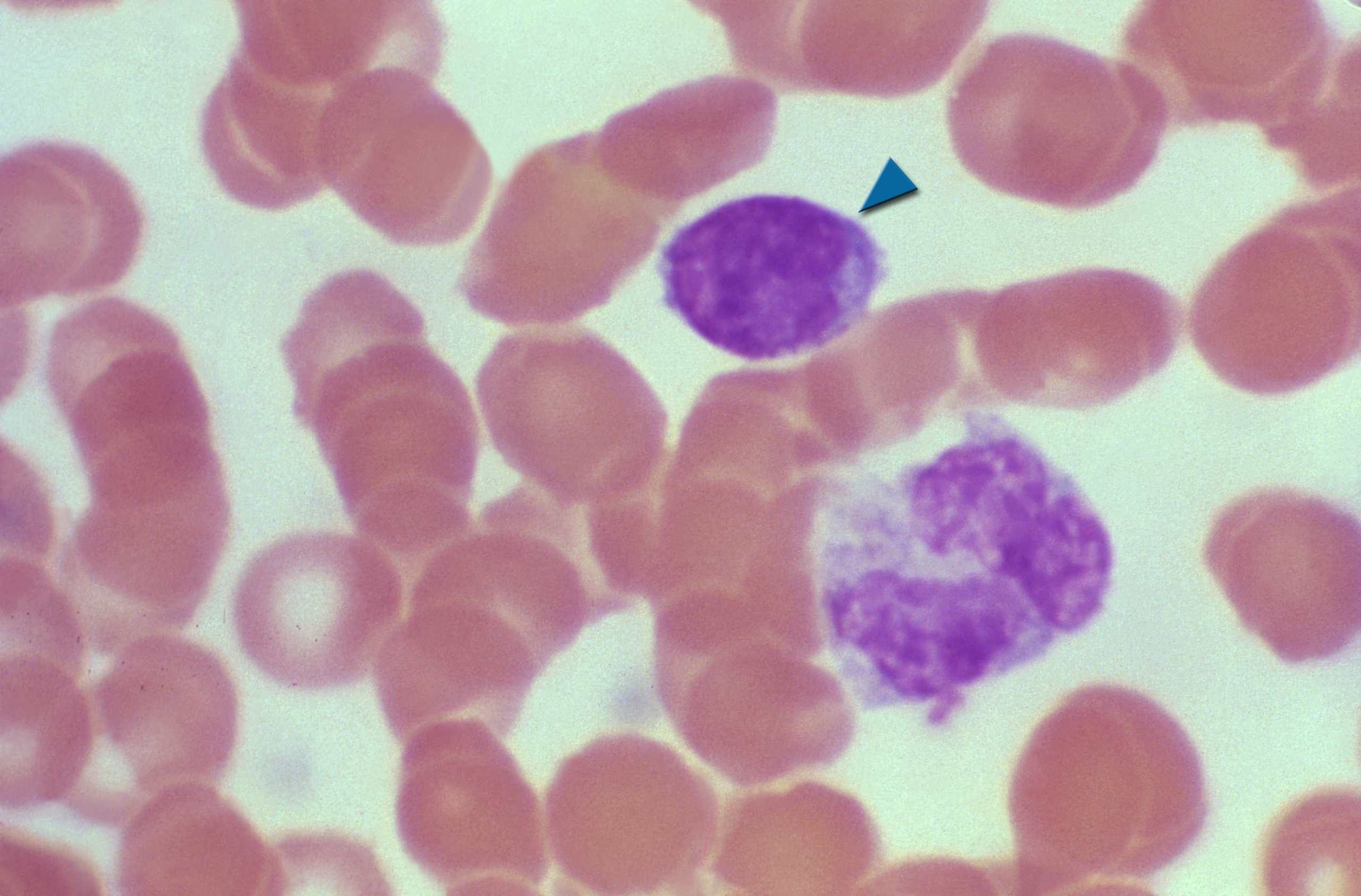
Lymphocytes are the cells behind antibodies and long-term immunity. Look for a small, round cell where a dark, round nucleus takes up almost all of the space, leaving a thin rim of pale blue cytoplasm.
Monocytes
Monocytes are the largest white blood cells. They leave the blood and become macrophages, which clean up debris and germs in the tissues. They have a folded, kidney- or horseshoe-shaped nucleus and a lot of gray-blue cytoplasm.
Eosinophils
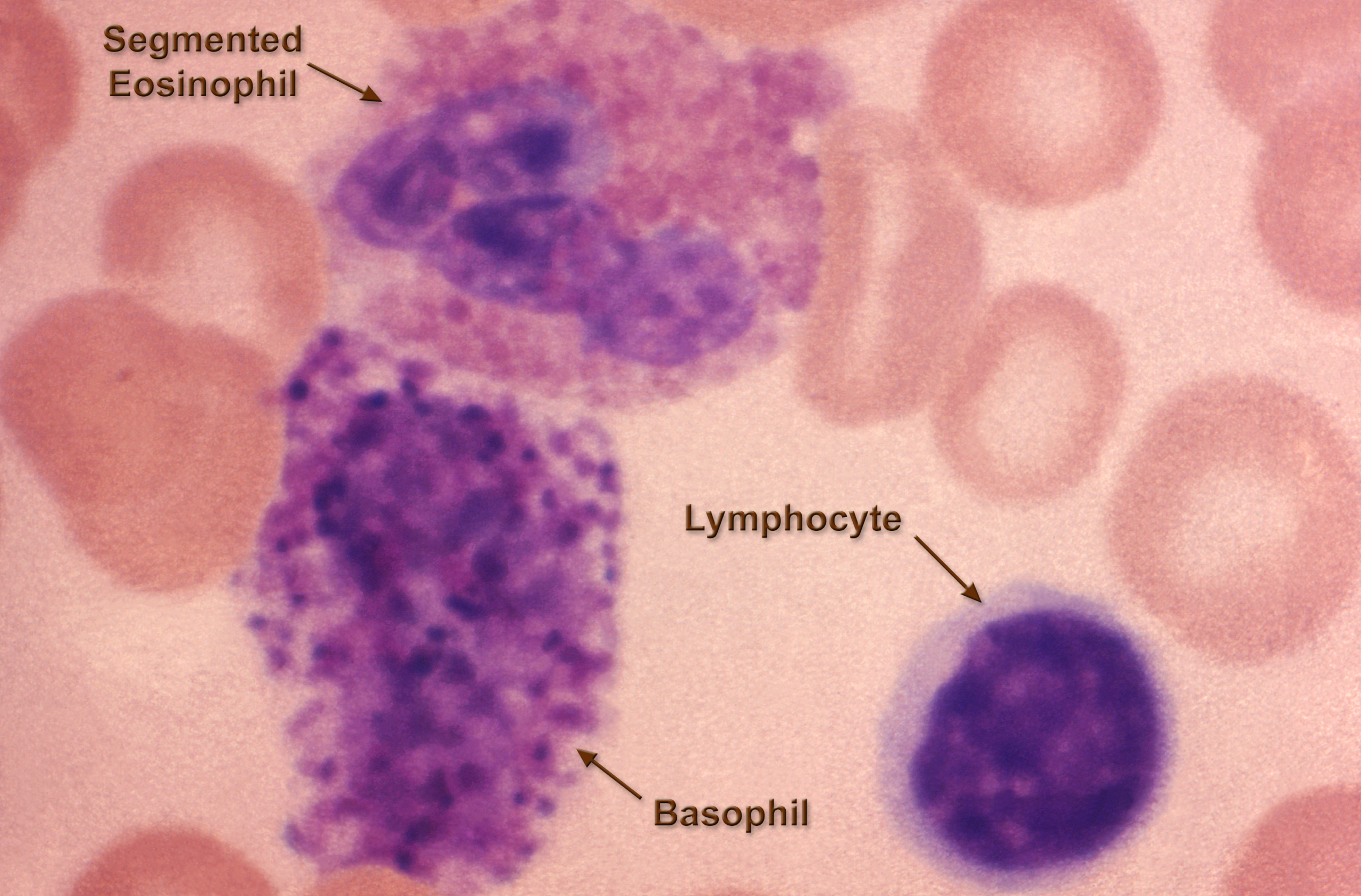
Eosinophils fight parasites, such as worms, that are too large to be engulfed, and they play a role in allergies and asthma. They’re unmistakable: a two-lobed nucleus surrounded by large, bright orange-red granules.
Basophils
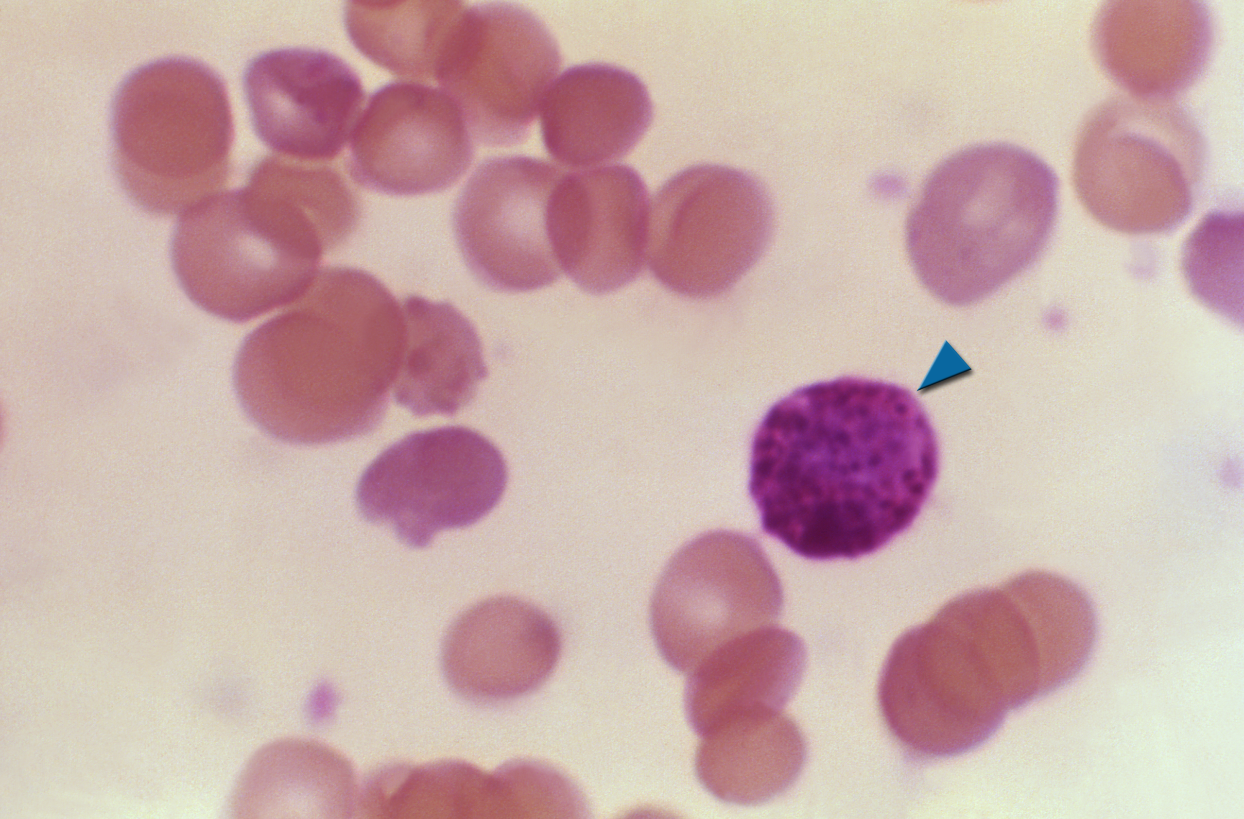
Basophils are the rarest white cells. They release histamine and other chemicals involved in allergic reactions and inflammation. Their dense, dark blue-purple granules are so heavy that they often hide the nucleus.
Platelets (thrombocytes)
Platelets aren’t full cells. They’re small fragments that break off from large cells in the bone marrow. On a stained slide, they appear as tiny purple specks, 2–3 µm across, scattered between the red cells, sometimes in small clumps.
Platelets help blood clot. When a blood vessel is damaged, they stick to the injury and to each other, forming a plug that slows bleeding. Too few platelets (thrombocytopenia) can cause easy bruising and heavy bleeding. Too many (thrombocytosis) is often a temporary reaction to infection, inflammation or injury, though some cases raise the risk of clots.
Plasma
Plasma is the liquid part of blood, about 55% of its volume. It’s a pale yellow fluid made mostly of water, with proteins, salts, nutrients, hormones and waste products dissolved in it. On a microscope slide, plasma isn’t visible as a separate structure; it’s simply the background. You can see it on its own by spinning blood in a centrifuge: the heavier cells sink, and the plasma settles on top as a clear yellow layer.
What abnormal blood looks like under a microscope
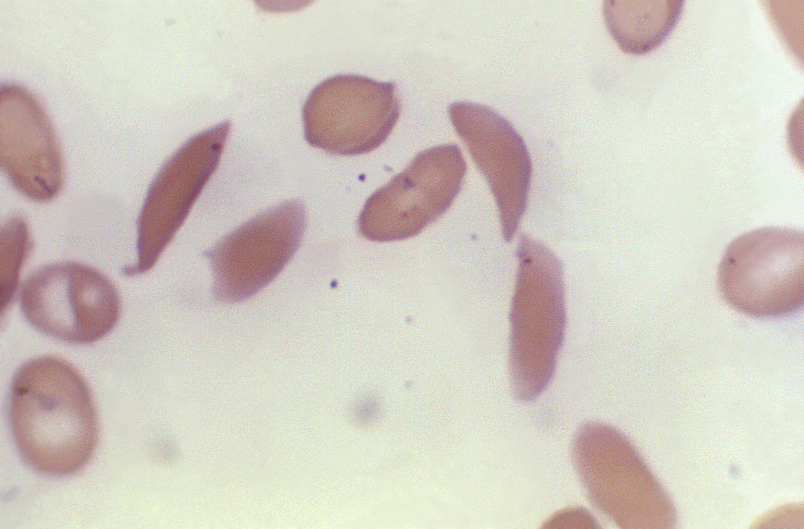
Doctors and lab scientists examine blood smears to spot disease. A few examples of what they look for:
- Sickle cell disease: some red cells are stiff and curved into a crescent or “sickle” shape instead of round discs.
- Iron deficiency anemia: red cells are smaller and paler than normal, with larger central pallor.
- Malaria: the parasites show up as small ring-shaped forms inside red blood cells on a Giemsa-stained smear.
- Megaloblastic anemia (from low vitamin B12 or folate): some neutrophils have six or more nuclear lobes.
- Abnormal counts: far too many or too few white cells can point to infection or to blood cancers such as leukemia.
This section is for education only. A home microscope can’t diagnose a blood condition; see a doctor if you have concerns about your health.
How animal blood looks different
Most animals’ blood has the same basic parts as ours, but there are some striking differences under the microscope.
- Nucleated red blood cells: human (and most mammal) red cells lose their nucleus, but birds, reptiles, amphibians and fish keep it. Their red cells look like oval discs with a dark nucleus in the middle.
- Oval red cells in mammals: camels and their relatives are the exception among mammals, with oval rather than round red cells.
- Different blood colors: octopuses and many other mollusks use copper-based hemocyanin instead of hemoglobin, which turns their blood blue when it carries oxygen. Some marine worms have green blood from a pigment called chlorocruorin. Certain skinks in New Guinea have green blood because of high levels of the bile pigment biliverdin. Antarctic icefish have no hemoglobin at all, and their blood is nearly colorless.
Frog blood vs. human blood
Frog blood is a popular comparison in biology classes. Frog red blood cells are much larger, oval-shaped and nucleated, so each one has a dark spot in the middle. Human red cells are smaller, round and have no nucleus. Both have white blood cells with nuclei. Instead of platelets, frogs have thrombocytes, small nucleated cells that do the same clotting job.
What is a blood smear?
A blood smear (also called a peripheral blood film) is a thin layer of blood spread on a slide, stained and examined under a microscope. Labs use it to check the size, shape and color of blood cells and to estimate how many there are. It can help diagnose anemias, infections, blood cancers and inherited conditions like sickle cell disease, as well as conditions such as polycythemia vera (too many red cells) and hemolytic uremic syndrome, in which red cells break apart.
How to look at blood under a microscope at home
The safe, easy option: prepared slides
The best way to start is with a prepared, stained blood smear slide. They’re sold individually or in sets of biology slides, and they show all the cells described above with no blood handling needed. They’re the best choice for kids and classrooms.
Making your own blood smear
If you want to make your own smear, use only your own blood, collected with a sterile, single-use lancet. Never handle anyone else’s blood, and dispose of lancets and slides safely.
You’ll need:
- A compound microscope with a 100x oil immersion objective (400x works for red cells)
- Clean glass slides (two per smear)
- A sterile single-use lancet and an alcohol wipe
- A quick blood stain kit (Wright-Giemsa or Leishman stain)
- Immersion oil
Steps:
- Clean your fingertip with an alcohol wipe, let it dry, and prick it with the lancet.
- Place a small drop of blood near one end of a clean slide.
- Hold a second slide at about a 30–45° angle in front of the drop, and pull it back until it touches the drop so the blood spreads along its edge.
- Push the second slide forward in one smooth, quick motion to spread a thin film. A good smear thins out to a feathered edge.
- Let the smear air-dry completely.
- Fix and stain it following the instructions on your stain kit.
- Rinse gently, let it dry, and place it on the microscope stage.
- Start at 40x to find the thin “feathered” area, where cells lie side by side, then move up to 100x and 400x.
- For white cell details and platelets, add a drop of immersion oil and switch to the 100x objective (1,000x total).
Want to photograph what you see? A microscope camera makes it easy to capture and share your slides.
Blood under a microscope FAQ
What magnification do you need to see blood cells? Red blood cells are visible from about 100x and clear at 400x. To see white cell nuclei in detail and individual platelets, use 1,000x with oil immersion.
Why do red blood cells look pink on a slide? The stain (usually eosin, part of Wright’s or Giemsa stain) binds to hemoglobin and colors it pink. Without stain, red cells look pale yellow-red.
Do you need to stain blood to see it under a microscope? You can see unstained red blood cells, but white blood cells are nearly invisible without stain, and you can’t tell the five types apart.
Why don’t red blood cells have a nucleus? Losing the nucleus as they mature leaves more room for hemoglobin and lets the cell form its flexible biconcave shape, which helps it squeeze through tiny blood vessels.
Can you see bacteria or viruses in blood with a home microscope? Bacteria are at the very limit of a home compound microscope at 1,000x, and they are rarely present in healthy blood. Viruses are far too small to see with any light microscope.
Can I see my blood type under a microscope? No. Blood type depends on molecules on the cell surface that are too small to see. It requires a blood-typing test.
Curious what else looks amazing up close? Try yeast under a microscope or our list of things to look at under a microscope.
One Response