
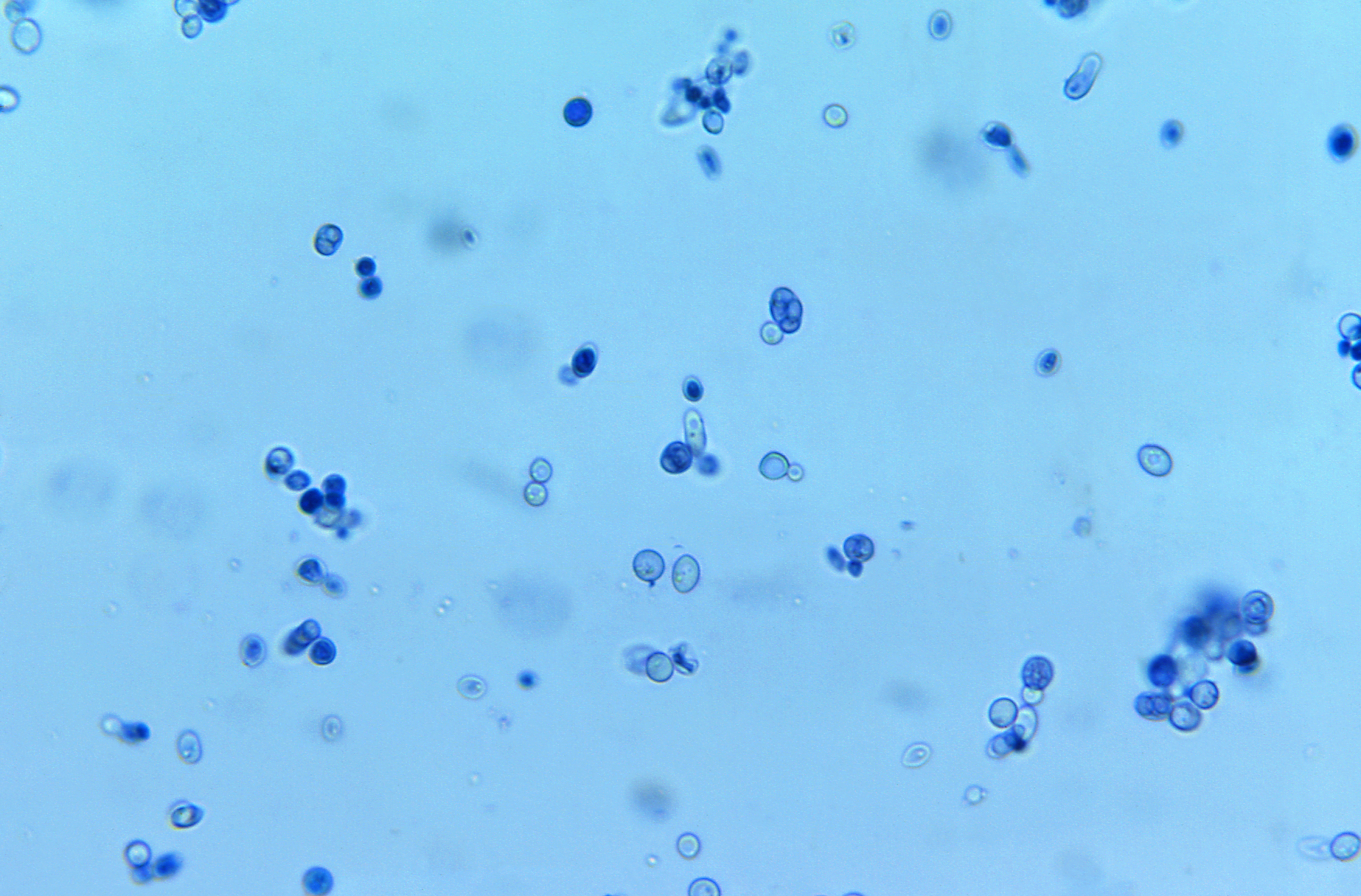
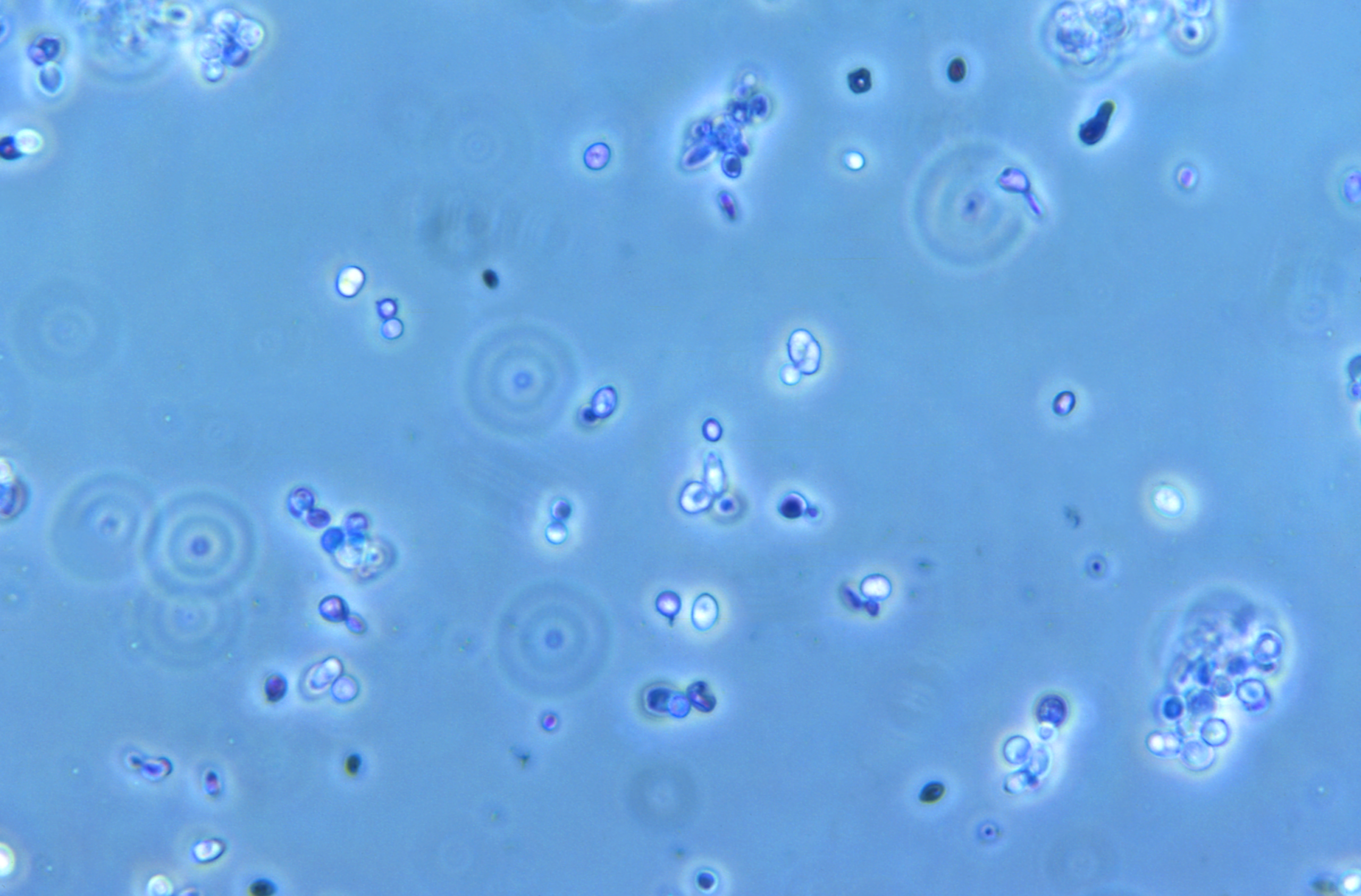
Under a microscope, yeast looks like a field of tiny round or oval cells, each about 3–10 µm across, scattered on their own, in pairs or in small clusters. At 400x you can see their shape clearly, and many cells have a small bump growing off one side. That bump is a bud, a new daughter cell forming, which is how yeast reproduces. Yeast cells are colorless unless stained and don’t swim or move on their own.
Yeast under a microscope: quick facts
What yeast looks like at different magnifications
*With a standard 10x eyepiece (field number 18). A 5 µm yeast cell takes up about 1% of the width of the view at 400x.
New to the controls? See our labeled guide to the parts of a microscope.
New to the controls? See our labeled guide to the parts of a microscope.
What is yeast?
Yeasts are single-celled fungi. Unlike bacteria, they’re eukaryotes: each cell has a nucleus and other internal compartments, just like plant and animal cells. About 1,500 species of yeast have been described, and they live almost everywhere, from soil and plant leaves to fruit skins and your own skin.
The word “yeast” comes from the Old English “gist” or “gyst” and an older Indo-European root meaning to boil, foam or bubble, a nod to the bubbles yeast makes as it ferments (Wikipedia). People have used yeast for thousands of years to make bread, beer and wine, long before anyone knew it was alive.
The yeast most people know is baker’s yeast, Saccharomyces cerevisiae, the same species used for brewing beer and making wine (University of Rochester). It’s also the easiest yeast to look at under a microscope, because you can buy it at any grocery store.
What yeast cells look like up close
- Shape: most yeast cells are round to oval. Depending on the species and growing conditions, they can also be elongated, cylindrical or lemon-shaped.
- Budding: look for cells with a small round bump attached. That’s a bud, a daughter cell growing off its parent. Eventually it pinches off and becomes a cell of its own. Sometimes buds stay attached and form short chains.
- Cell wall: each cell has a thin, clear outline, its cell wall.
- Vacuole: at 400x or 1,000x, many cells show a pale, clear area inside. That’s the vacuole, a storage compartment.
- Nucleus: yeast has a nucleus, but it’s very hard to see without special stains.
- No movement: yeast cells may drift in the liquid, but they can’t swim. If something is darting around on your slide, it isn’t yeast.
How to look at yeast under a microscope at home
Yeast is one of the easiest and most rewarding things to look at with a home microscope. You can make a slide in about 15 minutes with supplies from the grocery store.
You’ll need:
- A compound microscope (400x or more)
- Active dry baker’s yeast
- Warm water (not hot) and a pinch of sugar
- A glass slide, a coverslip and a dropper
- Optional: methylene blue stain (sold in pet stores as an aquarium treatment, or in science supply kits)
Steps:
- Stir about ¼ teaspoon of yeast and a pinch of sugar into half a cup of warm water.
- Wait 10–15 minutes, until it starts to foam. The yeast is now active and many cells will be budding.
- Put one small drop of the yeast mixture on the slide. If the liquid looks cloudy and thick, dilute it with a little more water first; too many cells will pile up.
- Lower a coverslip onto the drop at an angle to avoid air bubbles.
- Start at 40x to find the sample, then switch to 100x and 400x.
- Look for oval cells and buds. Try counting how many cells are budding.
Optional stained version: put a drop of methylene blue at one edge of the coverslip and touch a small piece of paper towel to the opposite edge. The paper towel draws the stain under the coverslip. Stained cells are easier to see.
The live/dead yeast test
Methylene blue also tells you which cells are alive. Living yeast cells break down the dye and stay colorless; dead cells can’t, so they turn blue. Brewers have used this test for decades to check whether their yeast is healthy. Try comparing fresh yeast with yeast that’s been mixed into very hot water.
Looking for more ideas? See our microscope activities for kids and things to look at under a microscope.
Ways scientists view yeast
- Brightfield microscopy: the standard method used by most home and school microscopes. Light passes up through the specimen from below. It shows cell shape, size and budding well, but unstained yeast is pale and low in contrast.
- Staining: dyes such as methylene blue or lactophenol cotton blue add color, so cells are easier to see, and some stains reveal whether cells are alive.
- Phase contrast microscopy: a special condenser and objectives turn tiny differences in the specimen into light and dark, making clear, unstained cells stand out without dye.
- Fluorescence microscopy: used in research labs. Cells are treated with fluorescent dyes or tagged proteins that glow under specific light, revealing structures like the nucleus, mitochondria and vacuoles in vivid color. It needs a dedicated fluorescence microscope.
Yeast vs. bacteria vs. mold under a microscope
Yeast vs. bacteria
The easiest clue is size. Bacteria are much smaller, usually 1–2 µm, and at 400x they look like tiny specks or short lines; you need 1,000x with oil immersion to see their shapes clearly. Yeast cells are several times bigger and easy to see at 400x. Yeast also has a nucleus, while bacteria don’t, and yeast never swims, while many bacteria can. Their cell walls differ too: yeast walls are made mainly of glucans and mannoproteins with some chitin, while bacterial walls are made of peptidoglycan. Learn more in what are microbes.
Yeast vs. mold and other fungi
Yeast is a fungus, but it lives as single cells. Molds and mushrooms are made of hyphae, long branching threads that form fuzzy networks. Under the microscope, that’s the giveaway: individual round cells mean yeast; long threads mean mold. Some fungi, called dimorphic fungi, can switch between a yeast form and a thread-like form depending on temperature and conditions. Read more about what fungi are and what they do.
Yeasts that cause infections, and how they look under the microscope
Most yeasts are harmless, and some live on and in our bodies normally. A few can cause infections, especially when the immune system is weakened. Lab scientists identify them partly by how they look under the microscope.
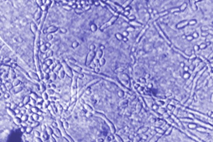
- Candida albicans lives in the mouth, gut and skin of most people without causing problems. When it overgrows, after antibiotics, with a weakened immune system or with high blood sugar, it can cause thrush and vaginal yeast infections. Under the microscope, Candida shows budding yeast cells along with long chains of stretched cells called pseudohyphae, a clue that separates it from baker’s yeast (Medical News Today).
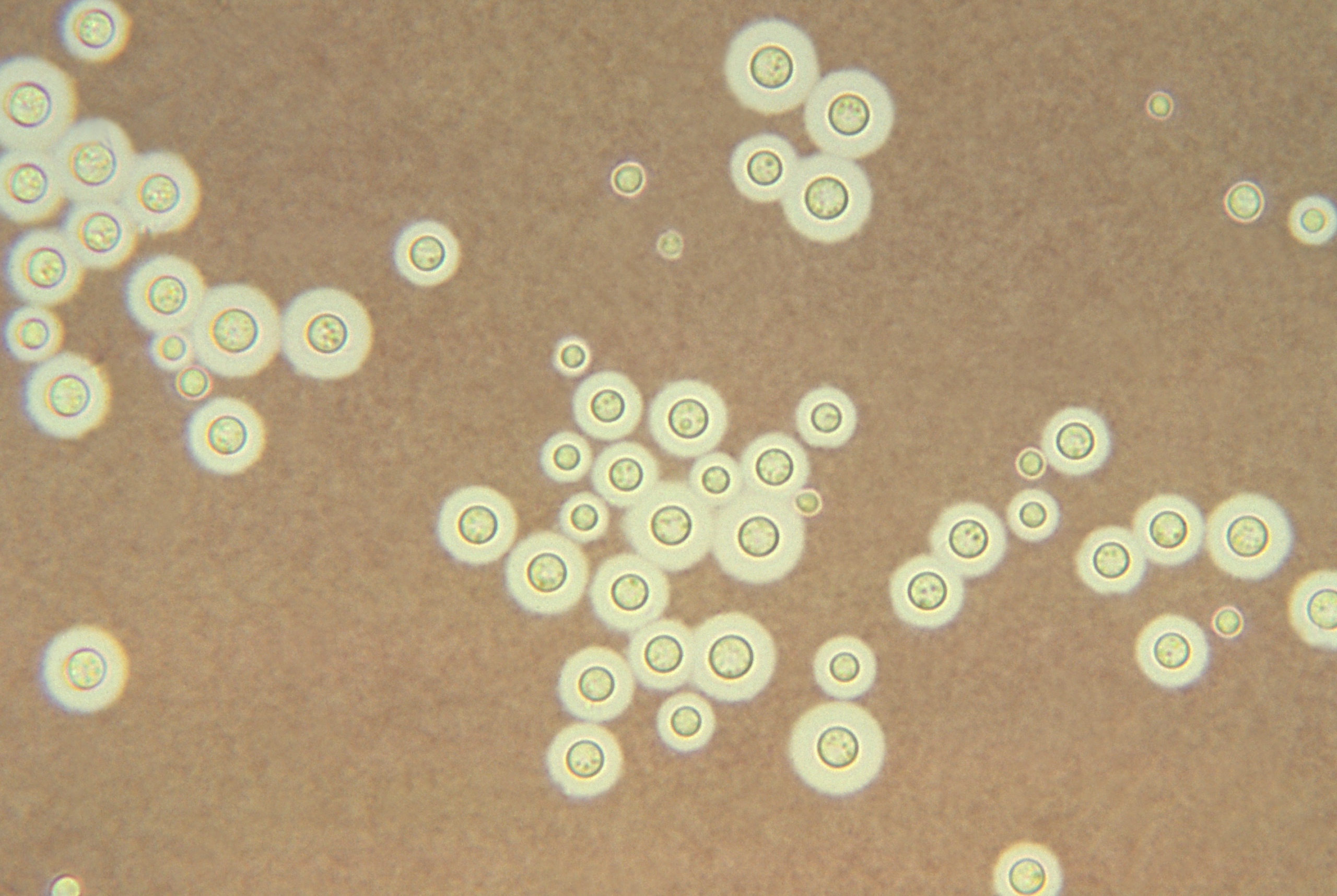
- Cryptococcus neoformans lives in soil and bird droppings and can cause serious lung and brain infections in people with weakened immune systems. It’s surrounded by a thick sugar capsule that helps it evade white blood cells. Under the microscope, an India ink stain makes that capsule show up as a clear halo around each cell.
- Malassezia yeasts live on everyone’s skin and are linked to dandruff, seborrheic dermatitis and tinea versicolor, a skin condition that causes patches of lighter or darker skin. Skin scrapings from tinea versicolor show a classic “spaghetti and meatballs” look: short threads mixed with round yeast cells.
This section is for education only. A home microscope can’t diagnose an infection; see a healthcare provider if you have symptoms.
What is yeast used for?
- Baking: yeast ferments sugars in dough and releases carbon dioxide, which makes bread and yeast-raised donuts rise.
- Beer and wine: yeast turns sugar into alcohol and carbon dioxide. Every beer and wine depends on it.
- Biofuel: yeast ferments sugar from crops like corn and sugarcane into ethanol, which is blended into gasoline.
- Food and nutrition: nutritional yeast, made from inactivated yeast, is a popular source of protein and B vitamins.
- Science: baker’s yeast is one of the most studied organisms on Earth. Its cells work much like human cells but are quick and cheap to grow, and in 1996 it became the first eukaryote to have its whole genome sequenced.
- Medicine: genetically engineered yeast is used to make medicines, including some insulin and hepatitis B vaccines. One yeast, Saccharomyces boulardii, is sold as a probiotic.
- Cosmetics: yeast extracts and proteins appear in some skin and hair care products.
When yeast causes problems in food
Some yeasts spoil food and drinks. Zygosaccharomyces species can grow in sugary, acidic foods like syrups, juices, jams and soft drinks that most microbes can’t tolerate, producing gas, off-flavors and cloudiness. In winemaking, Brettanomyces bruxellensis can create barnyard or medicinal smells that most winemakers consider a fault.
Yeast under a microscope FAQ
Can you see yeast with the naked eye? Not individual cells. The smallest thing the eye can see is about 0.1 mm (100 µm), and a yeast cell is roughly 20 times smaller. You can, however, see colonies of millions of yeast cells growing on bread or a lab plate.
What magnification do you need to see yeast? Yeast cells show up as dots at 100x and are clear at 400x, where you can see their shape and buds. Use 1,000x with oil immersion for more detail.
What does budding yeast look like? A larger round or oval cell with a smaller round bump attached to one side, like a snowman. The bump is a daughter cell that will eventually separate.
Is yeast alive? Yes. Dry baker’s yeast is dormant, but it becomes active within minutes in warm, sugary water. The methylene blue test above shows which cells are alive.
Can you see yeast with a pocket or phone microscope? Most pocket and clip-on phone microscopes magnify about 60–120x, which shows yeast as tiny specks at best. You need a compound microscope to see their shape.
Does yeast move under a microscope? No. Yeast cells have no flagella or cilia. They may drift with currents in the liquid, but they can’t swim.
Want to keep exploring? See what blood looks like under a microscope or learn how to grow mushrooms at home, yeast’s much bigger fungal cousins.
2 Responses